
Digital Radiography and Fluoroscopy Digital Radiography (x-rays) use a small amount of ionizing radiation to produce images of bones and tissue without requiring actual film. X-rays are useful for examining lungs and bone. Fluoroscopy is a type of x-ray imaging used to provide moving images that can be viewed via closed-circuit television monitor. Thus, the radiologist can view specific body parts while they are functioning. Fluoroscopy is especially valuable in evaluating the lower gastrointestinal system.
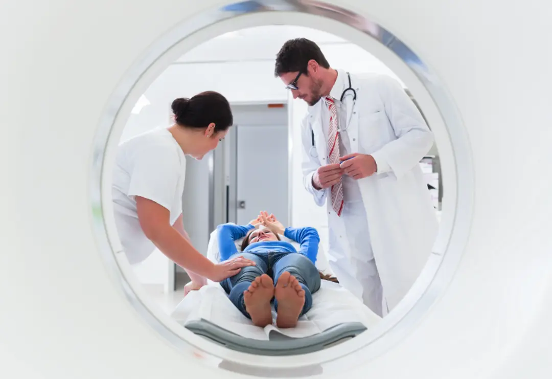
Providing Cutting-Edge Diagnostic Imaging and Intervention
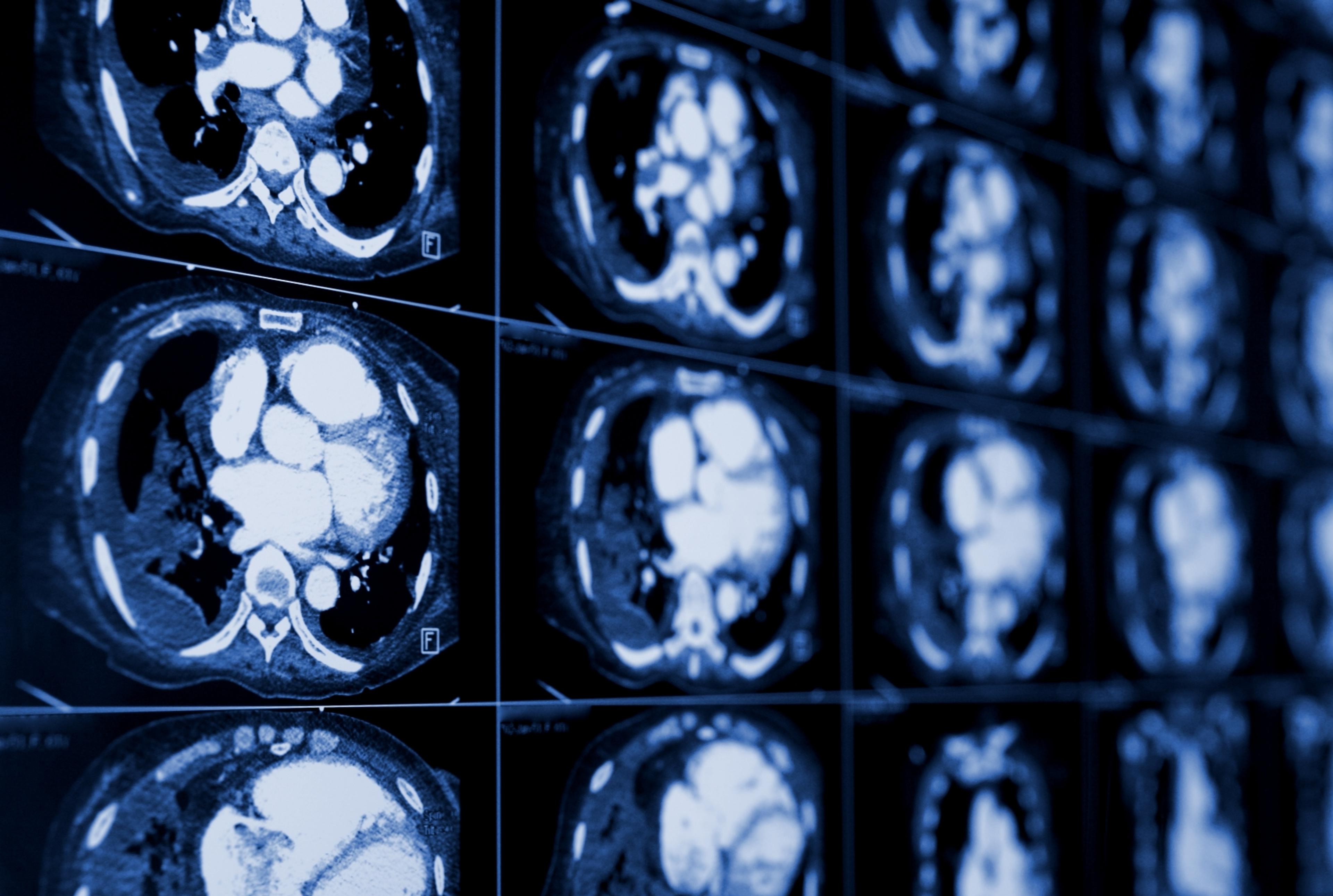
CT scanning joins specialized x-ray equipment with a computer to produce multiple images or pictures of the inside of the body. The images are joined together in cross-sectional views of the area studied, then examined on a computer monitor or printed.
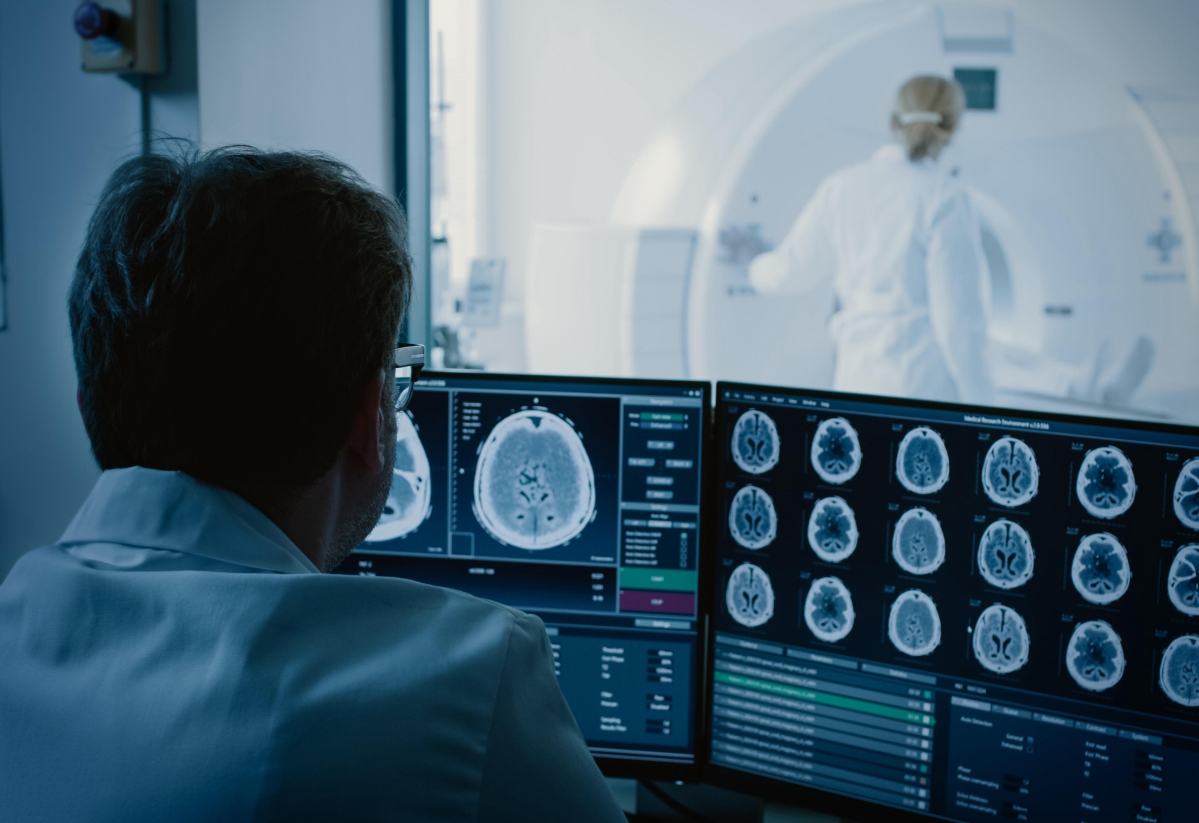
Providing Cutting-Edge Diagnostic Imaging and Intervention
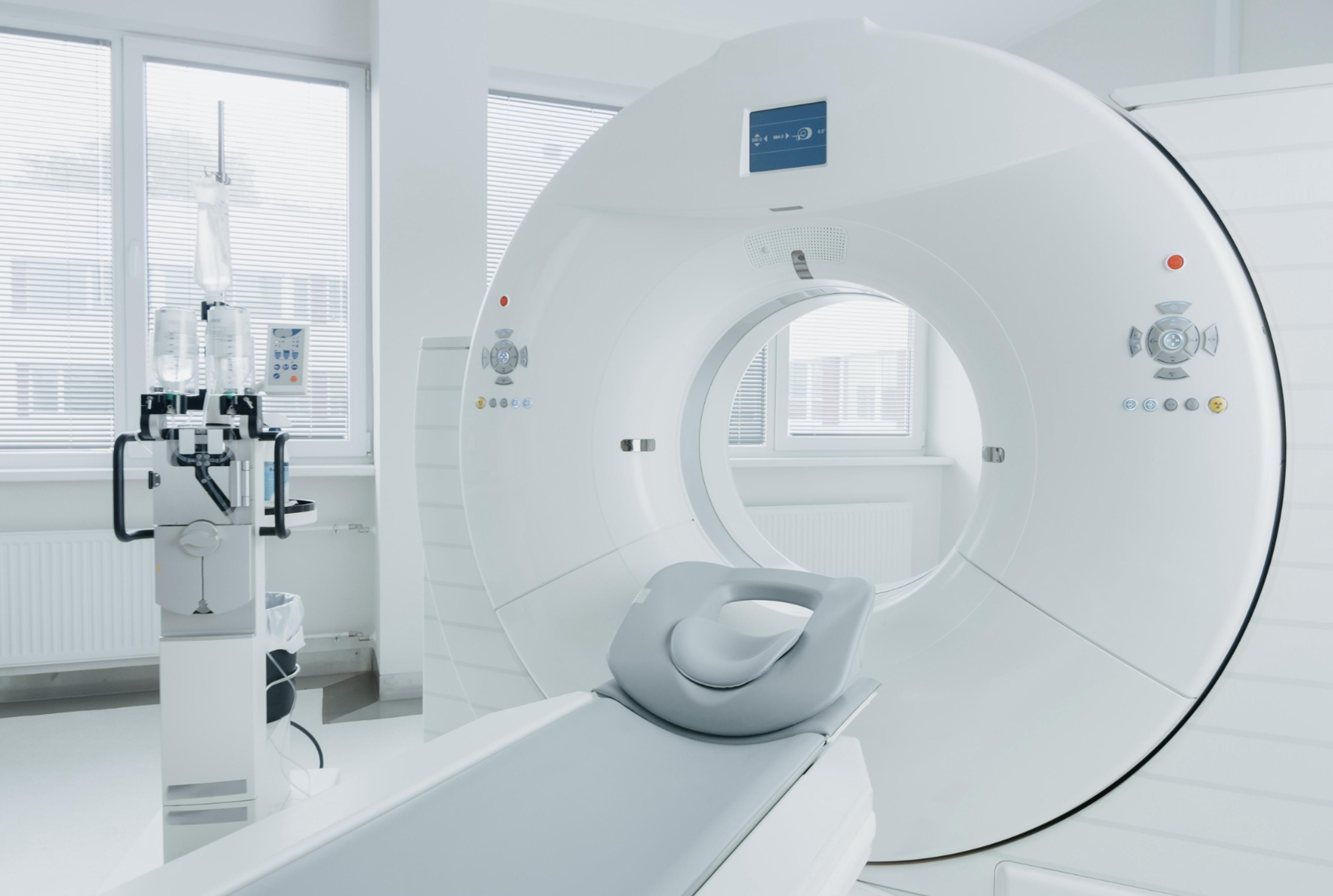
Using a large magnet, radio waves, and a computer, the MRI produces detailed cross-sectional images of organs, soft tissues, bone, and virtually all other internal body structures. Images are examined on a computer monitor or printed. MRI does not use x-rays.

Providing Cutting-Edge Diagnostic Imaging and Intervention
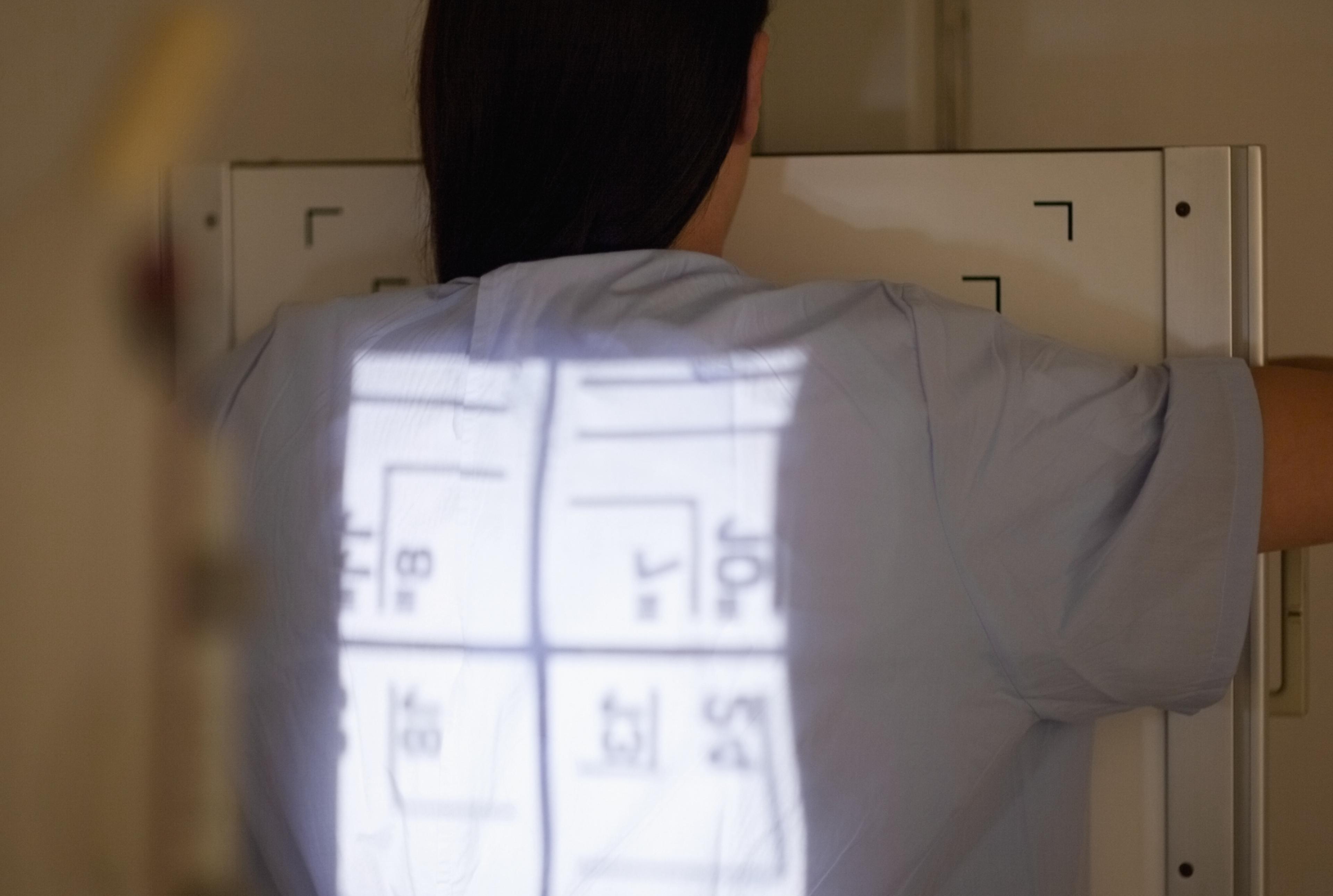
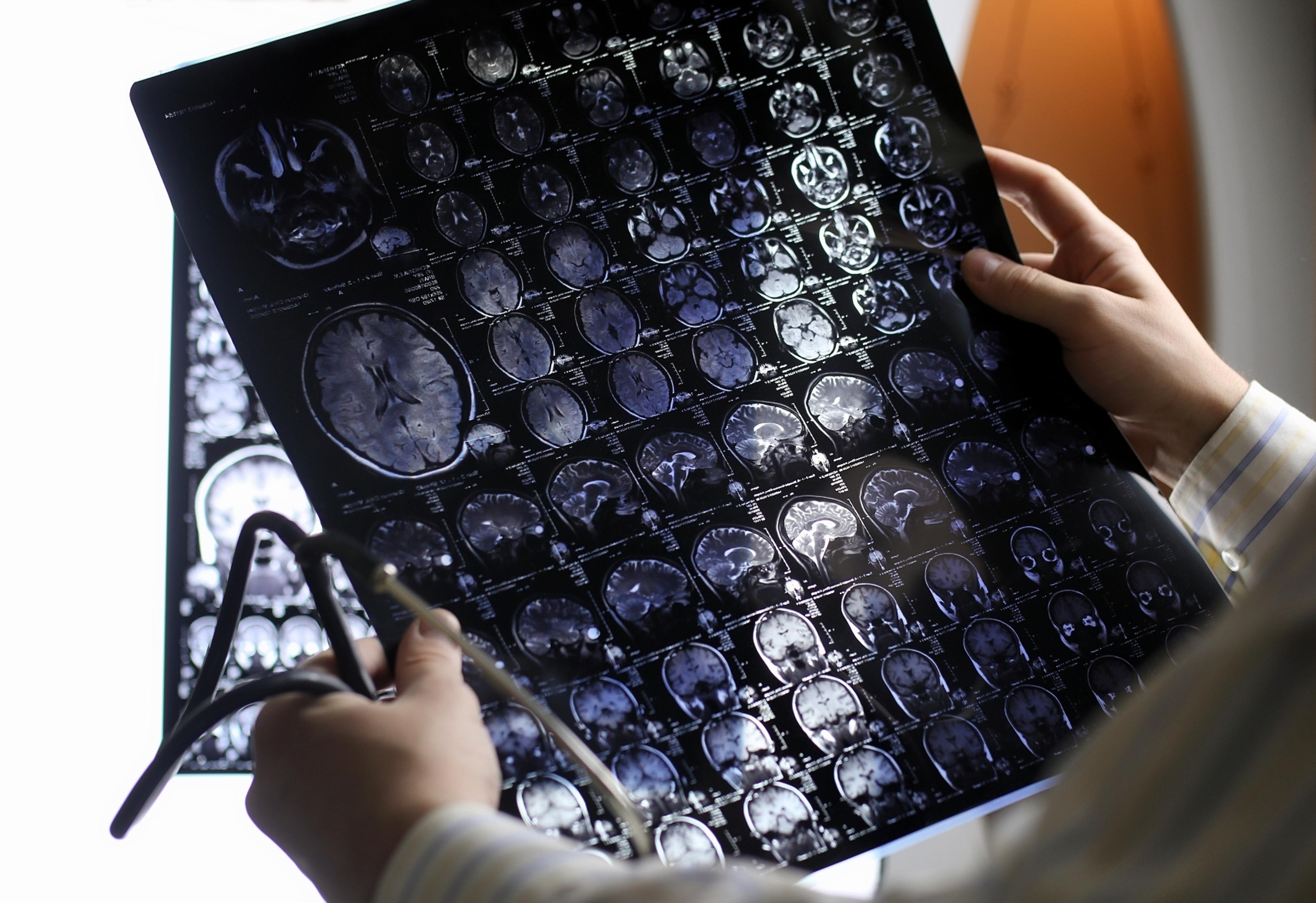
Nuclear Medicine utilizes a low risk amount of radioactive substance called a radionuclide, which is used to view the physiological function of the system being examined. This type of imaging is useful for detecting tumors, aneurysms, inadequate blood flow to various tissues, blood cell disorders and inadequate functioning of organs. Commons exams performed include imaging of the gallbladder and bile duct, thyroid gland, liver, kidneys and spleen, lungs and gastrointestinal tract.
Providing Cutting-Edge Diagnostic Imaging and Intervention
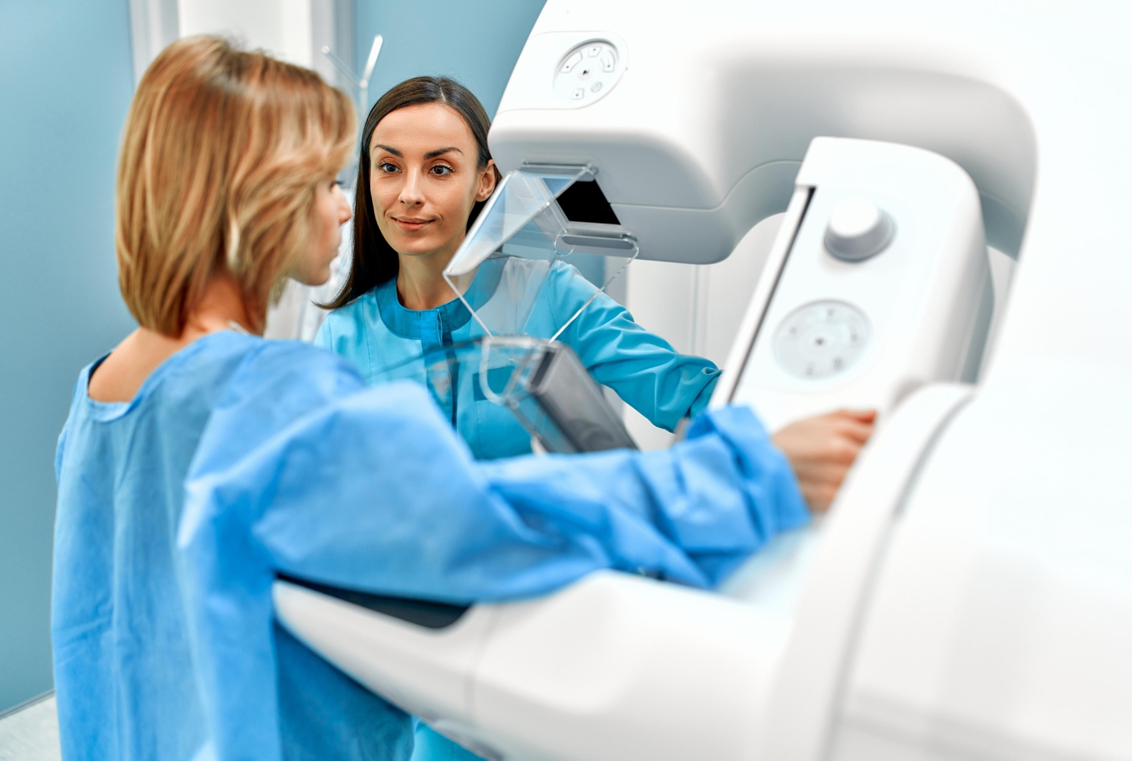
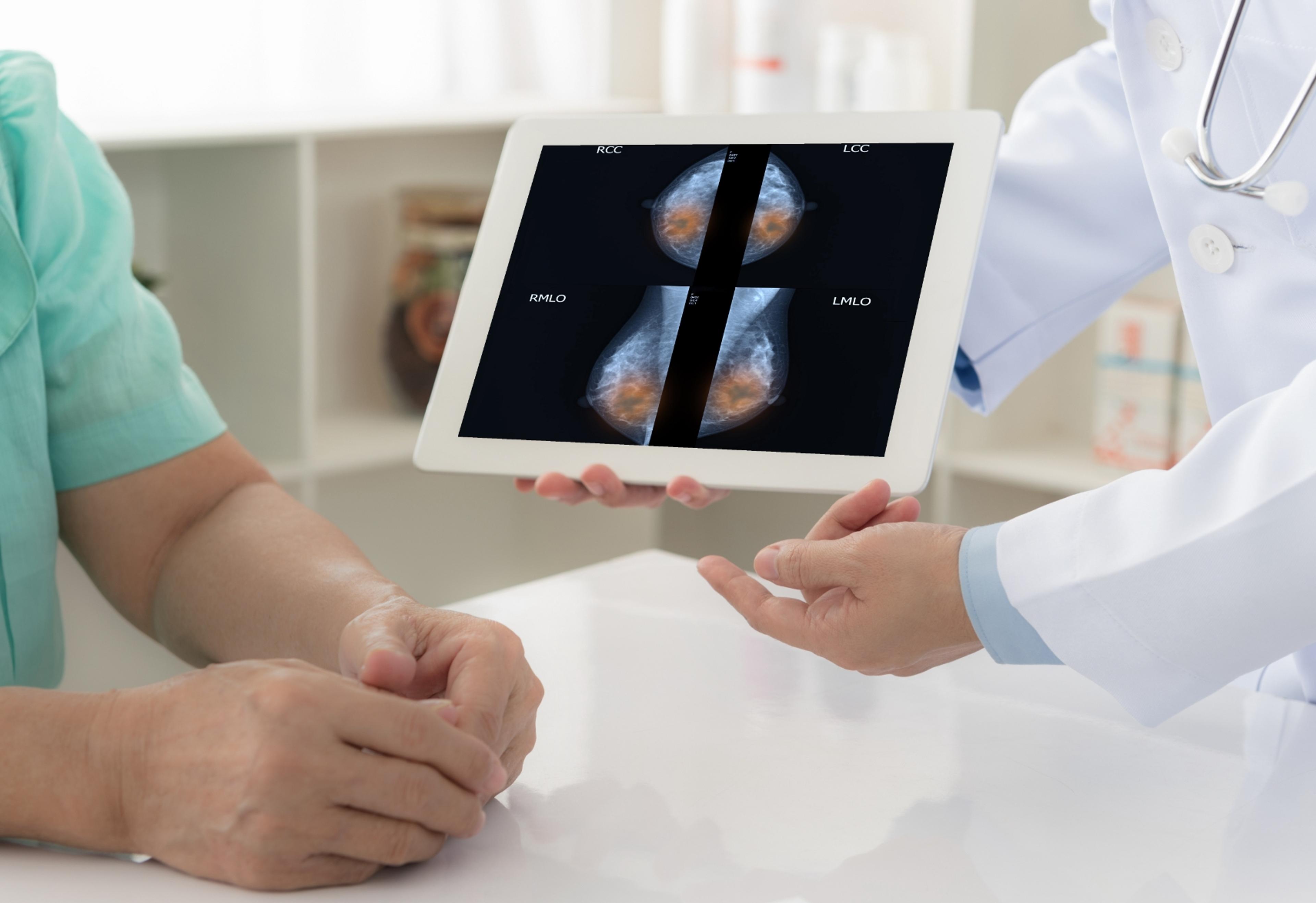
Digital mammography and CAD (Computer-Aided Detection), both relatively new technologies, are utilized to perform this simple x-ray examination of the breast(s). Mammography can detect cancer that can be too small to be felt or detected any other way.
Providing Cutting-Edge Diagnostic Imaging and Intervention

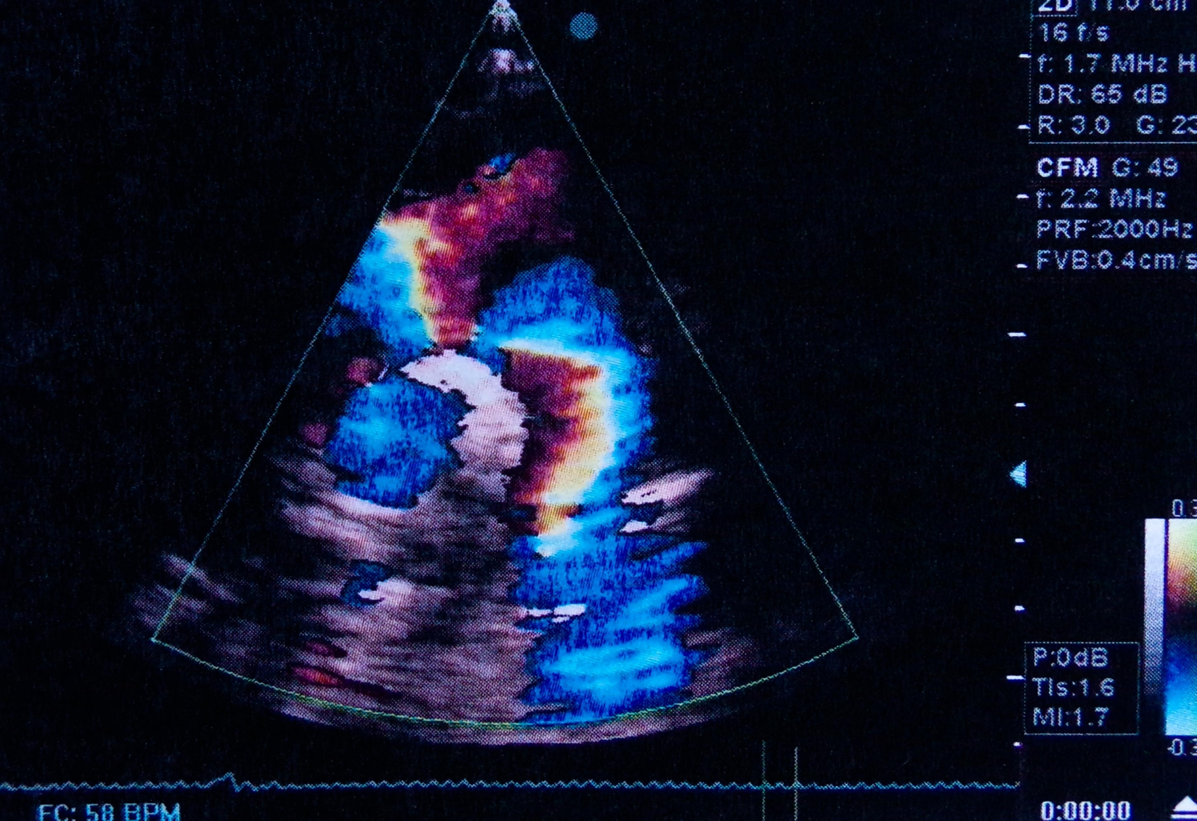
Ultrasound imaging uses high-frequency sound waves to produce pictures of the body's internal organs, movement within the organs, and blood flow through the vessels. Ultrasound exams do not use x-rays. Ultrasound images are captured in real-time.
Providing Cutting-Edge Diagnostic Imaging and Intervention

